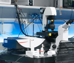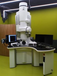
The JEM-F200 “F2” is a highly advanced state-of-the-art 200kV S/TEM electron microscope with cold field emission gun with high brightness and narrow energy spread. The microscope is equipped with cryo-tomography stage, hole-free phase plate, Fishione Model 2550 Cryo Transfer Tomography Holder, TVIPS TemCam – XF416 cooled CMOS camera, bright-field and dark-field STEM detectors, 100 μm2 windowless SDD EDS detector and a beryllium specimen holder. A new automated sample holder transfer system, the SpecPorter, facilitates sample loading. The microscope offers high performance, versatility and ease of use. The machine is pre-set at 120 and 200kV, thus allowing to collect EM data from specimen for which either high contrast or high resolution is preferred.
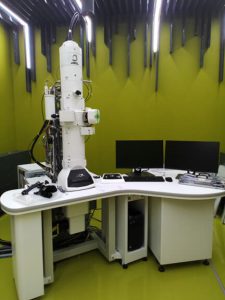
Up to 120 kV TEM with a tungsten or LaB6 filament electron beam source and a bottom-mounted FLASH 2kx2k CMOS camera. The machine is running at 120 kV, thus allowing to collect EM data from biological specimens for which high contrast is preferred to high resolution. A point resolution of 0.38 nm is available along with magnification range from 10x to 1.200.000x. The Limitless Panorama (LLP) software allows for automatic acquisition of large areas of the specimen at high resolution by image stitching. A software for correlative light and electron microscopy is also available. The microscope is very well suited for routine sample screening and acquisition of the micrographs in publication quality.
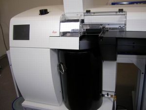
The Leica EM PACT2 is a high pressure freezing system for vitrifying samples up to 200 µm in thickness without the artefacts of chemical fixation. It preserves the biological samples in near-to-native state for further high-resolution EM observation. Bayonet loading device provides automatic specimen orientation. After freezing, the specimen is ejected into liquid nitrogen bath. The temperature/pressure curve is displayed for each run allowing the precise control of the process. Internal memory allows to collect up to 8 000 freezing runs. High-pressure frozen samples can be processed by either Frozen Hydrated Sectioning or Freeze Substitution. The Freeze Fracture Holder is also available.
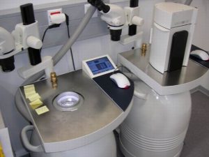
Leica EM AFS2 performs freeze substitution and progressive lowering of temperature, then allowing low temperature embedding and resin polymerization. It is equipped with UV lamp. Specimen can be visualized with the attached stereomicroscope under internal LED illumination. The temperature working range is from 140 °C to +70°C. The 35 l Dewar enables long protocols. Liquid nitrogen filling from outside of specimen chamber allows refilling during the run. The “Deep Freeze” function allows sample transfer at temperatures around -140 °C. Adjustable transfer function of gaseous (evaporated) liquid nitrogen excludes humidity and oxygen, which results in no contamination of the specimen. Several substitution systems are available: Substitution-Capsule and Flat Embedding System, New Microtube Embedding System EM AFS2 and Sapphire Disc FS System. The Leica EM FSP (freeze substitution processor) is an automatic reagent handling system. Mounted on the Leica EM AFS2, it dispenses reagents for both FS and PLT applications minimizing operators contact with toxic media and reducing risk to lose a specimen.
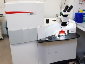
The Leica EM ICE is a high pressure freezing system for vitrifying samples up to 200 µm in thickness without the artefacts of chemical fixation. It preserves the biological samples in near-to-native state for further high-resolution EM observation. The EM ICE solution uses a unique, alcohol-free freezing principle to allow a superior cryo-fixation of the specimen enabling better quality results to be obtained. Quick sample loading and fully automated freezing process allows to minimize the manipulation time before freezing. After freezing, the specimen is ejected into liquid nitrogen bath. The temperature/pressure curve is displayed for each run allowing the precise control of the process. Synchronizing light stimulation (blue light 405 nm) and high pressure freezing enables to visualize highly dynamic processes and structural changes in photosensitive specimens with nanometer resolution and millisecond precision.
High-pressure frozen samples can be processed by Frozen Hydrated Sectioning, Freeze Substitution or Freeze Fracture.
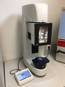
The EM GP2 plunge freezes fluid or extremely thin samples spread on an electron microscopy grid into liquid ethane after removing excess fluid by automatic blotting. Prior to freezing the sample is maintained in a temperature and humidity controlled environmental chamber, adjustable between +4°C and +60°C and room humidity to 99 %. It allows quick, safe and reproducible preparation of high-quality vitrified samples for single particle analysis or cryo – (S)TEM tomography research applications.
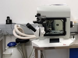
The Leica EM ACE900 is a high-end sample preparation system allowing to perform freeze fracture, freeze etching, and e-beam coating in one instrument. The high resolution carbon/Pt mix coatings by e-beam with rotating cryo stage offer flexible shadowing options for any TEM and SEM analysis. With a load lock for sample and knife transfer and gate valves for each e-beam source, the instrument can always stay under vacuum which ensures fast and clean working conditions. Freeze-fracture and replica immunolabeling technique is especially suitable for studies of membranes, bacteria, cell organelles.
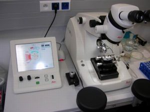
The Ultramicrotome Leica EM UC6 provides easy preparation of semi- and ultrathin sections as well as perfect, smooth surfaces of biological samples for TEM, SEM, AFM and LM examination. Eucentric movement of the stereomicroscope offers optimized positioning for approach with glass and diamond knives. It also helps in section observation at low water levels (e.g. for Lowycryls and dry sections) and cryosectioning without loss of ergonomic posture. Three independent brightness-controlled LED light sources provide illumination for toplight, backlight and specimen trans-illumination. The Integrated Antivibration System prevents influence from external vibrations. Motorized North-South and East-West movement of the knife stage in conjunction with the touch-sensitive control panel enables fast and safe alignment of knife and specimen with help files and prompts to hand for beginners (up to seven different user-settings can be stored). Programmable knife and cutting movements offer significant ease for trimming (Autotrim mode).
Low Temperature Sectioning System Leica EM FC6
Leica EM FC6 in combination with the Ultramicrotome Leica EM UC6 is designed for ultrathin cryosectioning of biological specimens in the range from -185 °C to -15 °C. Improved sectioning quality is provided with contact free through the wall specimen arm for vibration free sectioning, eucentric knife rotation with centre click stop from outside for easy alignment of knife and blockface, individual temperatures for specimen, knife and gas, automatic rapid cooling function to save time when cooling down to temperatures below -165°C. Specimen holder is locked with torque limited screw to provide optimum locking of sample without damage to the screw.
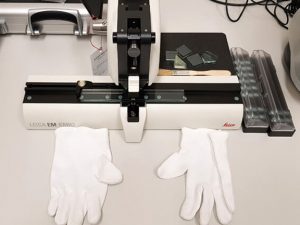
The balanced break method of the Leica EM KMR3 ensures perfect glass knives in three thicknesses 6,4 mm, 8 mm and 10 mm for semi-thin sections for EM and LM applications. The Leica EM KMR3 is easy to use and the automatic reset of breaking wheel and the scoring mechanism to “default” after a breaking cycle avoids handling errors.
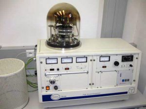
It enables transmission and scanning electron microscopy (TEM and SEM) specimen preparation, like producing carbon support films, preparation of replicas, shadowing, aperture cleaning, etc. The usage of a turbo molecular vacuum pump ensures oil free vacuum chamber environments. The further benefits are: simple operations, rapid pump down and high vacuum operation (routinely 10-5/10-6mbar). All accessory power supplies are vacuum interlocked for user protection and cannot be energized unless the chamber is under vacuum. Mounted on a 300 mm (12”) diameter stainless steel base plate, the TEM set includes resistive evaporation sources for 6 mm diameter carbon rods, metal evaporation (tungsten basket), tiltable specimen holder and base, glow discharge work chamber and baseplate electrode. The jigging is a one piece assembly making it easy to remove or modify. A “cool” sputtering head (not routinely mounted) allows SEM sputter coating. The base has 10 ports through which electrical, vacuum and water connections can be made. A rotating source shutter is also available. A 250 mm (10”) diameter bell jar provides the vacuum chamber.
Electron microscopy (EM) is the method of choice to study subcellular compartments in biological samples with high resolution. In many cases, the cellular compartments are defined by the content of specific molecules or by a presence of specific functions and they are morphologically inconspicuous in EM preparations. Such compartments can be detected using antibodies with attached label, usually gold particles. In this case, however, it is necessary to delineate the borders of the immunogold clusters in order to define the topology of the compartment. However, the labeling has a point pattern, while a compartment is a coherent area. In addition, some background labeling is always present which complicates identification of the labeled compartments. Stereological image analysis is therefore required to interpret the density distribution of gold particles.
More information – http://nucleus.img.cas.cz/gold/.

