Ústav molekulární genetiky AV ČR, v. v. i.

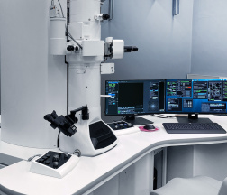
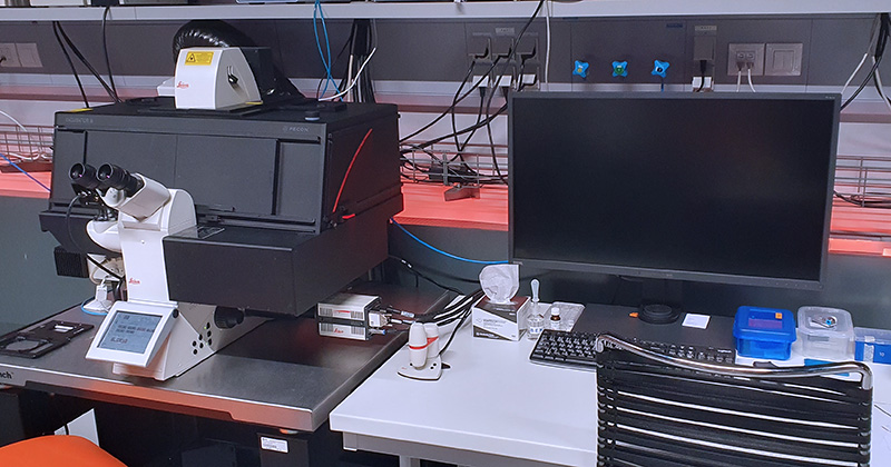
Inverted microscope DMi8 with confocal head Leica TCS SP8 is a confocal laser scanning microscope with full transmitted light equipment including transmitted light PMT and Nomarski contrast (DIC) available for all objectives. Leica TCS SP8 it is a dichroic mirror-based system, using interference filters for light filtering and beam-splitting. The confocal head is equipped with two fluorescence PMT detectors and three highly sensitive HyD detectors. The laser power is modulated by acousto-optical tunable filter (AOTF).
| Methods | Confocal scanning fluorescence imaging Transmitted light imaging Brightfield and Nomarski contrast (DIC) (manual) Tile-scans & merge in LAS X software Time-lapse experiments Multi-positions experiment Photo-kinetic experiment FRET (SE or Acceptor photobleaching) |
| Illumination | 405 nm diode laser, 50mW 488 nm solid-state laser, 20 mW 552 nm solid-state laser, 20 mW 638 nm solid-state laser, 30 mW HLX 100 W halogen lamp for transmitted light HXP 120W/45C VIS Hg lamp – Leica EL6000 for fluorescence |
| Objectives | HC PL FLUOTAR 10x/0.30 DRY; FWD 11.0 | BF, POL, DIC HC PL FLUOTAR 10x/0.30 DRY; FWD 11.0 | BF, POL, DIC HC PL APO 20x/0.75 IMM CORR CS2; FWD 0.66 | BF, POL HC PL APO 40x/1.30 OIL CS2; FWD 0.24; CG 0.17 | BF, POL, DIC HCX PL APO 63x/1.3 GLYC CORR 37°C; FWD 0.28; CG 0.14-0.18 | BF, POL HC PL APO 63x/1.40 OIL CS2; FWD 0.14; CG 0.17 | BF, POL, DIC |
| Eyepiece filtercubes | DAPI (A) (Ex: 360/40, Em: LP 425) FITC (I3) (Ex: 470/40; DM 510; Em: LP 515) RHOD (N2.1) (Ex: 537/45; DM 580; Em: LP 590) Cy5 (Y5) (Ex: 620/60; DM 660; Em: 700/75) CFP (Ex: 436/20; DM 455; Em: 480/40) |
| Detectors | 2x photomultiplier tube (PMT) 3x supersensitive hybrid detectors (HyD) 1x transmitted light detector |
| Confocal head | Acousto-optical tunable filter (AOTF) Low incident angle dichroic beam splitters Standard scanner (1-1800 Hz line frequency) Maximum scanner resolution 8192×8192 pixels Hardware zoom 0.75x-48x |
| Dichroic mirrors | 488/552/638 nm triple excitation dichroic 488/552 nm dual excitation dichroic Substrate RT 15/85 |
| Stage | Motorized microscope stage with Super Z-galvo scanning insert for fast and precise Z movement |
| Aditional equipment | Antivibration table |
| Software | LAS X |
| Location | room no. 0.172 (Green) |
| Phone | ext. 3172 |
| Booking | Calpendo („SP8“ ) |

The inverted microscope DMi8 with laser scanning confocal head Leica TCS SP8 is a confocal laser scanning microscope with full transmitted light equipment including transmitted light PMT and Nomarski contrast (DIC) available for all objectives. The confocal head is equipped with two fluorescence PMT detectors and three highly sensitive HyD detectors with time resolved gating function useful e.g. for removal of auto-fluorescence. The system has a wide-range white light laser with pulse picker (WLL PP) which allows for very versatile illumination tailored for any fluorophore. The combination of white light laser, AOBS and spectral sliders on detector cascade allows the fine tuning of excitation laser line selection and precise definition of emission spectra. Confocal head light path is equipped with optical image rotation. Stability for long-term live cell imaging experiments is ensured by a hardware autofocus system (AFC) and optional OKOLAB stage-top incubation chamber, which provides temperature, CO2, and humidity control.
| Methods | Confocal scanning fluorescence imaging Brightfield and Nomarski contrast (DIC) (automated) Tile-scans & merge in LAS X software Time-lapse experiments Multi-positions experiment |
| Illumination | Lamp for transmitted light: Leica EL6000 with HXP 120W/45C VIS Hg lamp – for fluorescence Excitation lasers: Pulsed white light laser (WLL2) – 470-640 nm with 1nm step, 8 parallel laser lines, 1.5 mW per each Continuous 405 nm DMOD Flexible laser – 50 mW |
| Objectives | HC PL FLUOTAR 10x/0.30 DRY; FWD 11.0 | BF, POL, DIC HC PL APO 20x/0.75 IMM CORR CS2; FWD 0.66 | BF, POL HC PL APO 40x/1.30 OIL CS2; FWD 0.24; CG 0.17 | BF, POL, DIC HC PL APO 63x/1.40 OIL CS2; FWD 0.14; CG 0.17 | BF, POL, DIC HC PL APO 100x/1.4 OIL STED WHITE CS2; FWD 0.13; CG 0.17 | BF, POL, DIC (OPTIONAL – UPON REQUEST) HC PL APO 86x/1.20 W CS2; FWD 0.30 mm| BF, POL, DIC |
| Eyepiece filtercubes | DAPI (A) (Ex: 360/40; DM 400; Em: LP 425) FITC (I3) (Ex: 470/40; DM 510; Em: LP 515) RHOD (N2.1) (Ex: 537/45; DM 580; Em: LP 590) Cy5 (Y5) (Ex: 620/60; DM 660; Em: 700/75) |
| Detectors | 2x photomultiplier tube (PMT) 400-800 nm 3x supersensitive hybrid detectors (HyD) 400-720 nm 1x transmitted light detector |
| Confocal head | Acousto-optical tunable filter (AOTF) Acousto-optical beam splitter (AOBS) Optical rotation Standard scanner (1-1800 Hz line frequency) Maximum scanner resolution 8192×8192 pixels Spectrally tunable detection Hardware zoom 0.75x-48x |
| Stage | Motorized microscope stage with Super Z-galvo scanning insert for fast and precise Z movement HW autofocus control |
| Aditional equipment | Antivibration table Incubation chamber Okolab (CO2, temperature, humidity) |
| Software | LAS X 64bit |
| Location | room no. 0.172 (Green) |
| Phone | ext. 3172 |
| Booking | Calpendo („SP8 WLL“ ) |
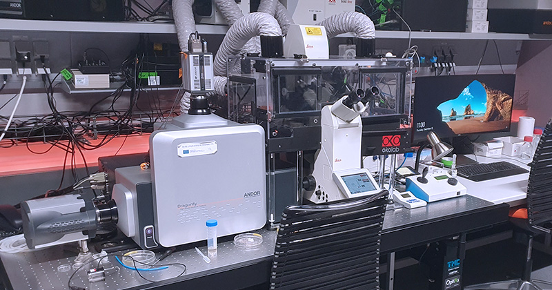
Dragonfly is a fully motorized multi-modal imaging platform with confocal and widefield modes. The core of the microscope is the confocal spinning disk, which combined with high-speed sCMOS and high-sensitive EMCCD cameras provides the system with exceptional speed and sensitivity. The low phototoxicity and photobleaching is ideal for live or delicate specimens. Dragonfly is 10-20 times faster than a traditional laser scanning confocal, which makes it optimal for imaging of fast dynamic events in live cell or for high throughput experiments. Photomanipulation experiments can be performed with Mosaic, a tool that allows for simultaneous illumination of multiple regions of interest of flexible shape and size. The semi-automated microinjector suited for adherent cells microinjection is also available. System is equipped with Super Resolution Radial Fluctuations super-resolution method.
| Methods | Confocal spinning disk fluorescence imaging Transmitted light imaging Brightfield and Nomarski contrast (DIC) Large tile-scans & merge in Fusion software Time-lapse experiments with HW autofocus Multi-positions experiment Photo-kinetic and opto-genetic experiment (limited software solution) Super Resolution Radial Fluctuations (SRRF) |
| Illumination | 405 nm solid-state laser, 200 mW 445 nm solid-state laser, 75 mW 488 nm solid-state laser, 150 mW 514 nm solid-state laser, 40 mW 561 nm solid-state laser, 100 mW 637 nm solid-state laser, 140 mW White LED source Leica DM for transmitted light CoolLED pE-300; 365 UV – white LED source for fluorescence 445 nm solid-state photo-manipulation laser for Mosaic, 1.3 W White LED source for Mosaic photo-manipulation (CoolLED pE-300; 365 UV) |
| Objectives | (HC PL APO 10x/0.40 DRY CS2; FWD 2.2; CG 0.17 | BF, POL, DIC) (OPTIONAL, UPON REQUEST) HC PL APO 20x/0.75 IMM CORR CS2; FWD 0.66 | BF, POL, DIC HC PL APO 40x/1.10 W CORR CS2; FWD 0.65; CG 0.14-0.18 | BF, POL, DIC HC PL APO 63x/ 1.20 W CORR CS2; FWD 0.3; CG 0.14-0.18 | BF, POL, DIC (OPTIONAL, UPON REQUEST) HCX PL APO 40x/1.25-0.75 OIL λBL; FWD 0.13; CG 0.17 | BF, POL HCX PL APO 63x/1.40-0.6 OIL λB; FWD 0.12; CG 0.17 | BF, POL, DIC HCX PL APO 63x/1.3 GLYC CORR 37°C; FWD 0.28; CG 0.14-0.18 | BF, POL HCX PL APO 100x/1.4-0.7 Oil CS; FWD 0.13; CG 0.17 | BF, POL, DIC |
| Dichroic mirrors | 405/488/561/640 nm Quad excitation dichroic 399-452/514/640 nm Triple excitation dichroic 405/488/561/685 nm Quad excitation dichroic |
| Eyepiece filtercubes | DAPI (Ex: 350/50; DC: 400; Em: 460/50) CFP/YFP (Ex: 435/25, 500/20; DC: 450; Em: 470/25, 535/20) FITC (Ex: 480/40; DC: 505; 527/30) RHOD (Ex: 546/10; DC: 560; 585/40) |
| Cameras | Zyla 4.2 PLUS sCMOS camera – 2048 x 2048 pix; 6,5 µm pixel iXon Ultra 888 EMCCD camera – 1024 x 1024 pix; 13 µm pixel |
| Dual camera switching mirror | LP 500 nm dichroic mirror (reflection to Zyla camera) LP 565 nm dichroic mirror (reflection to Zyla camera) 100% mirror (Zyla camera) 100% transmission dummy glass (iXon camera) |
| Zyla camera emission filter wheel | 450/50 nm bandpass filter (DAPI) 480/40 nm bandpass filter (CFP) 525/50 nm bandpass filter (GFP, A488, FITC) 540/30 nm bandpass filter (YFP) 600/50 nm bandpass filter (RFP, TRITC) 700/75 nm bandpass filter (Cy5) 405 to 445-515-561-730 quad emission filter (DAPI/CFP/YFP/mCherry/DyLight) Polarization filter |
| iXon camera emission filter wheel | 450/50 nm bandpass filter (DAPI) 480/40 nm bandpass filter (CFP) 525/50 nm bandpass filter (GFP, A488, FITC) 540/30 nm bandpass filter (YFP) 600/50 nm bandpass filter (RFP, TRITC) 620/60 nm bandpass filter (mCherry) 700/75 nm bandpass filter (Cy5) 405-488-561-640 quad emission filter (DAPI/GFP/RFP/Cy5) |
| Spare camera emission filter wheel | Polarization filter 725/40 nm bandpass filter 405-488-561-640 quad emission filter (DAPI/GFP/RFP/Cy5) 405 to 445-515-561-730 quad emission filter (DAPI/CFP/YFP/mCherry/DyLight) 445-515-561 tripple emission filter (CFP/YFP/RFP) 488-561 dual emission filter (GFP/RFP) 445-515 dual emission filter (CFP/YFP) |
| Microinjection | Microinjector FemtoJet joined with micromanipulator InjectMan NI 2 (Eppendorf) For volumes up to 100 pl Compressor to deliver the required pressure Integrated coarse and fine manipulator Work range: 20 mm per axis Automated and programmable axial injection movement |
| Aditional equipment | Okolab incubation unit including climate chamber (CO2, temperature, humidity) antivibration table photomanipulation module Mosaic (for FRAP, bleaching, uncaging, opto-genetics…) |
| Software | Fusion – acquisition software Imaris – visualization software Andor iQ3 – controlling Mosaic module |
| Location | room no. 0.171 (Orange) |
| Phone | ext. 3171 |
| Booking | Calpendo („Dragonfly“) |

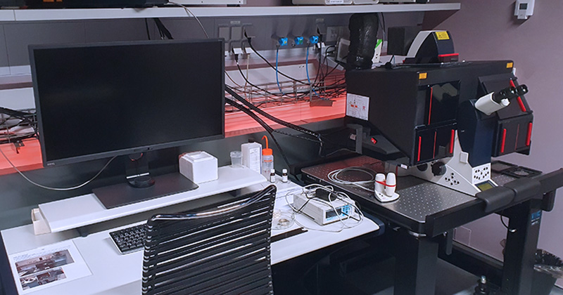
IXplore SpinSR SoRa is a fully motorized multimodal fluorescence microscope designed for gentle and rapid live-cell imaging. Built on the Evident IX83 platform, the system features a modular two-tier frame with an integrated focus adjustment for Köhler illumination and motorized objective turret that can accommodate up to six objectives. The system supports advanced imaging methods, including confocal spinning disk microscopy and super-resolution imaging based on optical reassignment technology. The system integrates cellSense software solution for multi-dimensional data acquisition, including z-stacks, multi-channel, tile-scans, time-lapse, and multi-position imaging. It also features scanR acquisition and analysis software for high-content screening. The microscope is equipped with drift focus correction (DFC) system to maintain z-axis stability during long term experiments.
| Methods | Confocal spinning disk with 50um pinholes (standard confocal) Confocal spinning disk with SoRa technology for enhanced resolution up to 120 nm Transmitted light imaging Large tile-scans & merge Time-lapse experiments with HW autofocus Multi-positions experiment DFC system based on IR laser for real-time imaging and precise Z-axis stabilization High-content screening with scanR software |
| Illumination | 405 nm solid-state laser, 50 mW 488 nm solid-state laser, 150 mW 561 nm solid-state laser, 100 mW 640 nm solid-state laser, 100 mW 785 nm solid-state laser, 100 mW Transmitted light: LED source IX3-LHLEDC CoolLED pE-300white for widefield fluorescence |
| Objectives | UPLXAPO 10X/0.40 DRY CS2; FWD 3.1 mm; CG 0.17 UPLXAPO 40X/0.95 IMM CORR CS2; FWD 0.18 mm; CG 0.11-0.23 UPLXAPO 60XO/1.42 OIL CS2; FWD 0.15 mm; CG 0.17 UPLXAPO 100XO/1.45 OIL CS2; FWD 0.13 mm; CG 0.17 |
| Dichroic mirrors | 405/488/561/640 nm Quad excitation dichroic 785 nm Single dichroic |
| Eyepiece filtercubes | DAPI (Ex: 350/50; DC: 400; Em: 460/50) FITC (Ex: 480/40; DC: 505; Em: 527/30) Cy3 (Ex: 546/10; DC: 560; Em: 585/40) |
| Cameras | ZHamamatsu ORCA-Fusion BT Type: sCMOS, global shutter Resolution: 2304 x 2304 pixels (5.2 MPix). Pixel Size: 6.5 x 6.5 µm Quantum Efficiency (QE): >95% at 550 nml |
| Dual camera switching mirror | LP 561 dichroic mirror: Directs emission to separate channels for simultaneous dual-camera imaging (e.g., GFP and RFP) Clear glass mirror: Allows full transmission to a single camera |
| Camera emission filter wheel | 2x 10-position wheels, each camera equipped with: DAPI (Ex: 405 nm) FITC (Ex: 488 nm) Cy3 (Ex: 561 nm) Cy5 (Ex: 640 nm) Cy7 (Ex: 785 nm) |
| Aditional equipment | Peacon incubation unit including climate chamber (CO2/O2, temperature, humidity) antivibration table |
| Software | CellSense acquisition software for confocal multimodal imaging ScanR Acquisition software for high-content screening ScanR Analysis software for high-content data analysis |
| Location | room no. 0.175 (Blue) |
| Phone | ext. 2750 |
| Booking | Calpendo („SpinSR“) |

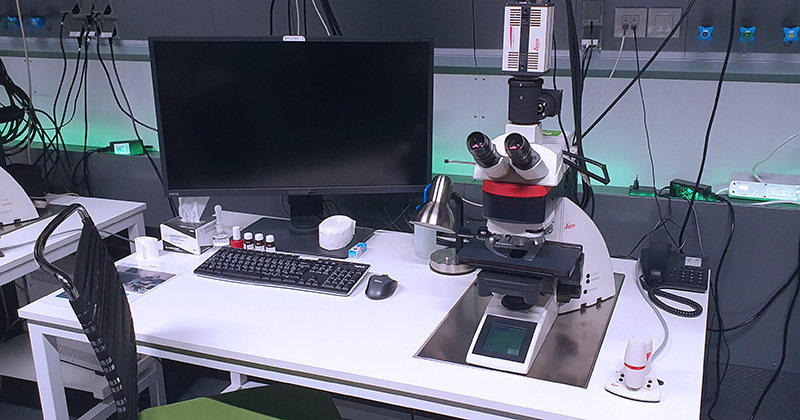
Upright routine widefield fluorescence microscope with motorized stage, fully automated transmitted and fluorescence light axis, and both monochromatic and color cameras. The monochromatic one is a high resolution sensitive grayscale sCMOS camera. The LAS X Navigator tool allows to create high-resolution image tile-scans of large areas using the precise motorized stage.
| Methods | Routine fluorescence imaging True color RGB imaging in transmitted light mode Brightfield/Darkfield and Phase contrast Large tile-scans & auto merge in LAS X Navigator |
| Illumination | HLX 100 W Halogen lamp for transmitted light HXP 120W/45C VIS Hg lamp – Leica EL6000 for fluorescence |
| Objectives | HC PL FLUOTAR 5x/0.15 DRY; FWD 12 | BF, DF, POL HC PL APO 10x/0.40 DRY PH1; FWD 2.2; CG 0.17 | BF, DF, POL, PH HC PLAN APO 20x/0.70 DRY PH2; FWD 0.59; CG 0.17 | BF, DF, POL, PH HCX PL APO 40x/0.75 DRY PH2; CG 0.17 | BF, DF, POL, PH HCX PL APO 63x/1.40 OIL PH3 CS; FWD 0.1; CG 0.17 | BF, POL, PH HCX PL APO 100x/1.40-0.70 OIL; FWD 0.09; CG 0.17 | BF, DF, POL |
| Filtercubes | DAPI (A4) (Ex: 360/40; DM 400; Em: 470/40) FITC (L5) (Ex: 480/40; DM 505; Em: 527/30) TRITC (N3) (Ex: 546/12; DM 565; Em: 600/40) Cy5 (Y5)(Ex: 620/60; DM 660; Em: 700/75) |
| Cameras | Leica K3C – color CCD camera, 2,4 μm pixel Leica DFC 9000 – monochromatic sCMOS camera; 6,5 µm pixel, QE: min. 82 % |
| Stage | Motorized microscope stage xyz |
| Software | LAS X, 64 Bit |
| Location | room no. 0.172 (Green) |
| Phone | ext. 3172 |
| Booking | Calpendo („DM6000“ ) |
Upright widefield fluorescence microscope with motorized stage, fully automated transmitted and fluorescence light axis and high-resolution sensitive monochromatic 16-bit sCMOS camera. System enables image rotation by software controlled camera rotator.
| Methods | Routine fluorescence imaging Brightfield/Darkfield and Phase contrast |
| Illumination | HLX 100 W Halogen lamp for transmitted light HXP 120W/45C VIS Hg lamp – Leica EL6000 for fluorescence |
| Objectives | HC PL FLUOTAR 6.3x/0.13 DRY; FWD 12.860 | BF, POL HC PL FLUOTAR 10x/0.30 DRY PH1; FWD 11.0; CG 0.17 | BF, DF, POL, PH HC PL FLUOTAR 20x/0.40 DRY PH1; FWD 7.5; CG 0.17 | BF, DF, POL, PH HC PLAN APO 20x/0.70 DRY PH2; FWD 0.59; CG 0.17 | BF, DF, POL, PH HCX PL APO 40x/0.75 DRY PH2; FWD 0.28 CG 0.17 | BF, DF, POL, PH HCX PL APO 63x/1.40 OIL; FWD 0.14; CG 0.17 | BF, POL, PH HCX PL FLUOTAR 100x/1.3 OIL PH3; FWD 0.13; CG 0.17 | BF, POL |
| Filtercubes | DAPI (A) (Ex: 360/40; DM 400; Em: LP 425) GFP (Ex: 470/40; DM 495; Em: 525/50) TRITC (N3) (Ex: 546/12; DM 565; Em: 600/40) Cy5 (Y5)(Ex: 620/60; DM 660; Em: 700/75) |
| Cameras | Leica DFC 9000 – monochromatic sCMOS camera; 6,5 µm pixel, QE: min. 82 % |
| Stage | Motorized microscope stage xyz |
| Software | LAS X, 64 Bit |
| Additional equipment | Electronic Camera rotator allows 360° physical rotation |
| Location | room no. 0.172 (Green) |
| Phone | ext. 3172 |
| Booking | Calpendo („DM6000-2“) |
Inverted widefield fluorescence microscope with motorized stage and fully automated transmitted and fluorescence light axis. The system is dedicated for live cell imaging. The stability of long-term time-lapse experiments with live cells is ensured by hardware autofocus system and environmental chamber for temperature, CO2 and humidity control. The microscope is also equipped with infinity port laser-scanner for photo-kinetic experiments (FRAP, iFRAP, Photo-activation). All the features are fully implemented in LAS X software. The LAS X Navigator tool allows to create high-resolution image tile-scans of large areas using the precise motorized stage.
| Methods | Routine fluorescence imaging True color RGB imaging in transmitted light mode Brightfield/Darkfield, Phase contrast, Modulation contrast Large tile-scans & auto merge in LAS X Navigator Time-lapse experiments with HW autofocus Multi-positions experiment Photo-kinetic experiments Large tile-scans & auto merge in LAS X Navigator |
| Illumination | LED light source for transmitted light HXP 120W/45C VIS Hg lamp – Leica EL6000 for fluorescence lasers for photomanipulation: – 405 nm laser (50 mW) – 488 nm laser (50 mW) |
| Objectives | HC PL FLUOTAR 5x/0.25, FWD 12 | BF, POL, PH, IMC N PLAN 10x/0.25 DRY; FWD 17.6 | BF, POL, PH, IMC HCX PL FLUOTAR L 20x/0.40 DRY CORR; FWD 6.9; CG 0-2 | BF, POL, PH, IMC HCX PL FLUOTAR 40x/0.95 DRY CORR; FWD 3.3; CG 0-2 | BF, POL, PH, IMC HCX PL APO 40x/1.25 oil | BF, POL HCX PL APO 63x/1.40-0.60 OIL; FWD 0.1; CG 0.17 | BF, POLBF, POL |
| Filtercubes | DAPI (A4) (Ex: 350/50; DM 400; Em: 460/50) FITC (Ex: 480/40; DM 505; Em: 527/30) TRITC (Ex: 546/10; DM 560; Em: 585/40) Y5 (Cy5) (Ex: 620/60; DM 660; Em: 700/75) |
| Cameras | Leica K3C – color CCD camera, 2,4 μm pixel Leica DFC 9000 – monochromatic sCMOS camera; 6,5 µm pixel, QE: min. 82 % |
| Stage | Motorized microscope stage xyz |
| Aditional equipment | PEACON incubation unit (CO2, temperature, humidity) |
| Software | LAS X |
| Location | room no. 0.171 (Orange) |
| Phone | ext. 3171 |
| Booking | Calpendo („DMI 8“ ) |
Inverted widefield fluorescence microscope DMi6000 with fully automated transmitted and fluorescence light axis. The transmitted light equipment includes differential interference contrast (DIC) prisms. An incubation chamber makes the system suitable for live-cell imaging. The total internal reflection fluorescence illumination module (Leica AM TIRF MC) controls the angle of the excitation beam and makes use of the total reflection to excite only molecules in the thin section in contact with the cover glass.
| Methods | Routine fluorescence imaging Brightfield, Nomarski contrast (DIC) Multi-positions experiment Time-lapse experiments TIRF microscopy experiments |
| Illumination | HLX 100 W Halogen lamp for transmitted light HXP 120W/45C VIS Hg lamp – Leica EL6000 for fluorescence solid-state TIRF laser 405 nm solid-state TIRF laser 488 nm solid-state TIRF laser 561 nm solid-state TIRF laser 633 nm |
| Objectives | HCX PL FLUOTAR 10x/0.30 DRY PH1; FWD 11.0 | BF, POL HCX PL FLUOTAR L 20x/0.40 CORR DRY PH1; FWD 6.9; CG 0-2 | BF, POL N PLAN L 40x/0.55 CORR DRY; FWD 3.3; CG 0-2 | BF, POL HCX PL APO 63x/1.3 GLYC CORR 37°C; FWD 0.28; CG 0.14-0.18 | BF, POL, DIC HCX PL APO 63x/1.40-0,60 OIL CS; FWD 0.1; CG 0.17 | BF, POL, DIC HCX PL APO 100x/1.46 OIL CORR CS; FWD 0.09; CG 0.1-0.22 | BF, POL, DIC |
| Filtercubes | A (Ex: 360/40; DM 400; Em: LP425) CFP-T (Ex: 422/44; DM 455; Em: 480/40) GFP-T (Ex: 490/20; DM500; Em: 525/50) YFP-T (Ex: 490/20, Em: 535/30) Cy3-T (Ex: 560/10, Em: 610/65) Cy5-T (Ex: 635/10, Em: 720/60) TRI (B/G/R) (ExFW: 420/30; 495/15; 570/20, Em: 457/22;530/20;628/28) FRET kostka pro CFP/YFP (ExFW: 436/12, 500/20; EmFW: 467/37, 545/45) QUAD-T (ExFW: 405/12, 490/20, 560/30,635/20; EmFW: 450/50, 525/36,600/32,705/72) |
| Cameras | Leica DFC350FX R2 – monochromatic CCD camera; 6,4 µm pixel Andor iXon 897 – high sensitivity EMCCD back-iluminated camera, 512×512 px, suitable for single molecule live cell imaging; 16 µm pixel |
| Stage | Motorized microscope stage with Super Z-galvo scanning insert for fast and precise Z movement |
| Aditional equipment | Incubation unit (CO2, temperature, humidity) Acousto-optical tunable filter (AOTF) – laser control |
| Software | LAS AF |
| Location | room no. 0.171 (Orange) |
| Phone | ext. 3171 |
| Booking | Calpendo („TIRF“) |
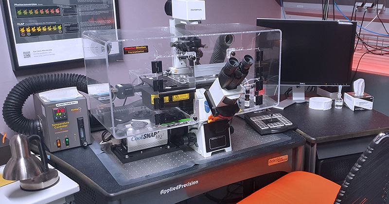
The system is built on the inverted widefield Olympus microscope version IX-71. The system is equipped with the HW autofocus system and an incubation chamber, and is suitable for multi-location and time-lapse experiments. The lasers for photomanipulation enable photokinetic experiments (FRAP) and FRET. The sensitive camera makes the microscope suitable for live cell imaging and low fluorescence intensity applications.
| Methods | Routine fluorescence imaging Brightfield, Nomarski contrast (DIC) Tile-scans Multi-positions experiment Time-lapse experiments with HW autofocus Photo-kinetic experiments |
| Illumination | Lumencore Spectra X LED light source for fluorescence lamp for transmitted light Lasers for photomanipulation (adjustable spot size): – 405 nm laser PoinSource iFlex2000 – 488 nm laser Coherent Sapphire (20 mW) |
| Objectives | U PLAN FL 20x/0.50 DRY PH1; FWD 1.6; CG 0.17 | BF, POL U PLAN FL 40x/0.75 DRY; FWD 0.51; CG 0.17 | BF, POL U APO/340 40x/1.35-0.65 CORR OIL; FWD 0.1; CG 0.17 | BF, POL PLAN APO N 60x/1.42 OIL; FWD 0.15; CG 0.17 | BF, POL, DIC |
| Exciation LED source | DAPI 390/18 CFP 438/24 GFP/FITC 475/28 YFP 513/17 RD-TR-PE 542/27 mCherry/Alexa594 575/25 Cy5 632/22 |
| Dichroic mirrors | Standard (DAPI, FITC, RD-TR-PE, Cy5) Live Cell (CFP, YFP, mCherry) Alexa (GFP, mCherry, Alexa 594) |
| Emission filter wheel | DAPI (435/48) FITC (523/36) RD-TR-PE (594/45) Alexa 594 (632/60) Cy5 (676/34) GFP (525/50) mCherry (632/60) CFP (470/24) YFP (559/38) |
| Eyepiece filers | The same as on emission filter wheel (excluding Cy5 filter) |
| Cameras | Photometrics CoolSANP HQ – high sensitive CCD camera, 1024×1024 px; 6,45 µm pixel |
| Stage | Motorized stage with xyz movement equipped with HW AF |
| Aditional equipment | Incubation unit (CO2, temperature, humidity) Antivibration table Hardware Ultimate autofocus for maintenance in-focus stability within the entire specimen |
| Software | SoftWorx – execution and evaluation 3D imaging – photo-kinetics experiments – time-lapse – FRET experiments – evaluation of co-localization – deconvolution |
| Location | room no. 0.171 (Orange) |
| Phone | ext. 3171 |
| Booking | Calpendo („DVcore“) |

Zeiss Axiom Zoom.V16 – Fully motorized fluorescence macroscope with high resolution objectives is equipped with two cameras and a motorized 16:1 zoom with apochromatic correction. The system is well suited for a wide range of bigger samples and has superior brightness in large object fields. ApoTome.2 uses structured illumination to improve contrast and to increase resolution of the optical sections.
| Methods | Fluorescence imaging on widefield OR Apotome (optical sectioning) mode Transmitted and/or reflected light True color RGB imaging in transmitted light mode Brightfield/Darkfield, Opaque contrast Large tile-scans & auto merge in ZEN Blue software Time-lapse experiments Multi-positions experiment |
| Illumination | HXP 120 V – metal halid lamp for fluorescence CL 9000 LED CAN – transmitted light source CL 9000 LED CAN – incident light source |
| Objectives | PlanNeoFluar Z 1.0x/0.25; FWD 56 | BF, DF, oblique illumination + Reflected light PlanNeoFluar Z 2.3x/0.57; FWD 10.6 | BF, DF, oblique illumination + Reflected light |
| Fluorescence filtercubes | DAPI Filter set 49 (Ex: G 365; BS FT 395; Em: BP 445/50) eGFP Filter set 38 HE (Ex: BP 470/40; BS FT 495; Em: BP 525/50) Cy3 Filter set 63 HE (Ex: BP 572/25; BS FT 590; Em: BP 629/62) Cy5 Filter set 50 (Ex: BP 640/30; BS FT 660; Em: BP 690/50) |
| Cameras | ZEISS Axiocam 305 – color CMOS camera; 3,45 µm pixel ZEISS Axiocam 512 mono – monochromatic CCD camera; 3,1 µm pixel |
| Additional equipment | ApoTome.2 for optical sectioning |
| Software | ZEN Blue 2.3 |
| Location | room no. 0.175 (Blue) |
| Phone | ext. 2750 |
| Booking | Calpendo („Apotome“) |
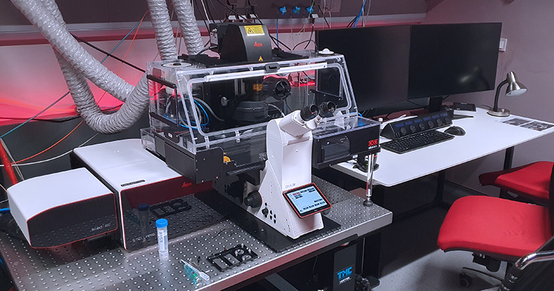
Fully motorized inverted microscope DMi8 with confocal head Leica STELLARIS 8 equipped with wide-range white light laser with pulse picker (WLL PP), highly sensitive time-resolved detection, FCS, and STED is a top-of-the-line system for a variety of high-end applications. The system is equipped with several acquisition modules for photo-kinetic experiments, FLIM or FLIM-FRET based experiments, the high-resolution imaging followed with Lightning adaptive deconvolution module, and FCS and FCCS modules for studying molecular dynamics. Furthermore, it contains a STED module with a TauSTED Xtend which can reach normal STED resolution with much lower depletion laser power, making it uniquely suited for super-resolution acquisitions of living cells. The system supports AI-based workflow for fast rare event detection using Autonomous Microscopy powered by Aivia software.
The combination of white light laser, AOBS and spectral sliders on detector cascade allows the fine tuning of excitation laser line selection and precise definition of emission spectra.
Stability of long-term live cell imaging experiments is ensured by a hardware autofocus system (AFC) and OKOLAB incubation system, which provides temperature, CO2, and humidity control.
| Methods | TauSTED Xtend – gentle fluorescence lifetime-based STED Confocal scanning fluorescence imaging with Lightning super-resolution module Transmitted light imaging Brightfield and Nomarski contrast (DIC) (automated) Tile-scans & merge in LAS X software Time-lapse experiments Multi-positions experiments Photo-kinetic experiments FRET (SE or Acceptor photobleaching) FLIM, FRET-FLIM experiments Rare event detection and Autonomous microscopy using Aivia software |
| Illumination | Excitation lasers: 405 nm CW diode laser, AOTF modulated, 100mW 440 – 790 nm Pulsed white light laser with pulse picker (WLL PP): – Range of wavelengths selection: 440 – 790 nm – Laser line selection step: 1 nm – Laser power:> 0,9 mW 440-485 nm,> 1,8 mW 485-790 nm – Possible laser frequencies: 80, 40, 20, 10, 5, 2,5 MHz – 8 channel AOTF – WLL PP in combination with AOBS allows up to 8 independent parallel laser lines Depletion lasers: – 589 nm pulsed STED, >1.5 W – 775 nm pulsed STED, >1.5 W 12V 100W LED source for transmitted light HXP 120W/45C VIS Hg lamp – Leica EL6000 for fluorescence |
| Objectives | HC PL FLUOTAR 10x/0.30 DRY CS2; FWD 11.0 mm | BF, POL, DIC HC PL APO 20x/0.75 IMM CORR CS2; FWD 0.67 mm | BF, POL HC PL APO 93x/1,30 GLYC motCORR STED WHITE; FWD 0.30 mm| BF, POL, DIC PL APO 40x/1.30 OIL CS2; FWD 0.24; CG 0.17 | BF, POL, DIC HC PL APO 63x/1.40 OIL CS2; FWD 0.14 MM| BF, POL, DIC HC HC PL APO 100x/1.4 OIL STED WHITE CS2; FWD 0.13; CG 0.17 | BF, POL, DIC (OPTIONAL – UPON REQUEST) |
| Eyepiece filtercubes | DAPI (Ex: 355/56, DC: 400, Em: 460/50) GFP (Ex: 470/40; DC 495; Em: 525/50) RHOD (Ex: 546/10; DC 560; Em: 585/40) CFP/YFP (Ex: 435/25 + 500/20; DC 450 + 515; Em: 470/25 + 535/30) |
| Detectors | 2x supersensitive silicon-based hybrid detectors (HyD-S) 2x supersensitive hybrid detectors (HyD-X) for very fast time-resolve acquisition (FLIM) 1x supersensitive hybrid detector (HyD-R) for long wavelengths (700 – 850 nm) 1x transmitted light TL – PMT detector |
| Confocal head | 8-channels acousto-optical tunable filter (AOTF) for fast laser intensity modulation Acousto-optical beam splitter (AOBS) Optical rotation Conventional linear scanner (1-2600 Hz line frequency) Maximum scanner resolution 8192×8192 pixels Hardware zoom 0.75x-48x Scanned field size 22 mm (diagonally) Resonant scanner (8000 Hz line frequency) Maximum scanner resolution 2496 x 2496 pixels Hardware zoom 1.25x-48x |
| Time-resolve modules | Tau-sense modules for time-resolved acquisition (time-resolved imaging without further analysis): – TauGating – TauContrast – TauSeparation STELLARIS FALCON: FAST Lifetime CONtrast (FALCON) is solution for fast image acquisition, processing and FLIM analysis with modules: – FLIM component fitting – FLIM Phasor diagram – FLIM-FRET analysis |
| Computational super-resolution module | Lightning – a super-resolution module based on mutual acquisition control and adaptive deconvolution based on SNR decision mask estimation. Maximal declared resolution is 120 nm laterally and 200 nm axially. |
| Stage | Motorized microscope stage with Super Z-galvo scanning insert for fast and precise Z movement |
| Additional equipment | Antivibration table OKO-LAB incubation unit (CO2 + O2, temperature and humidity control system) |
| Software | All features are fully implemented to the LAS X software LAS X modules: LAS X LiveDataMode LAS X 3D Visualisation LAS X Phasor LAS X FRAP, FRAP Zoomer LAS X Assay (Navigator) LAS X Lightning LAS X Navigator Expert LAS X Rare Event Detection Aivia |
| Location | room no. 0.174 (Red) |
| Phone | ext. 2433 |
| Booking | Calpendo („Stellaris“) |
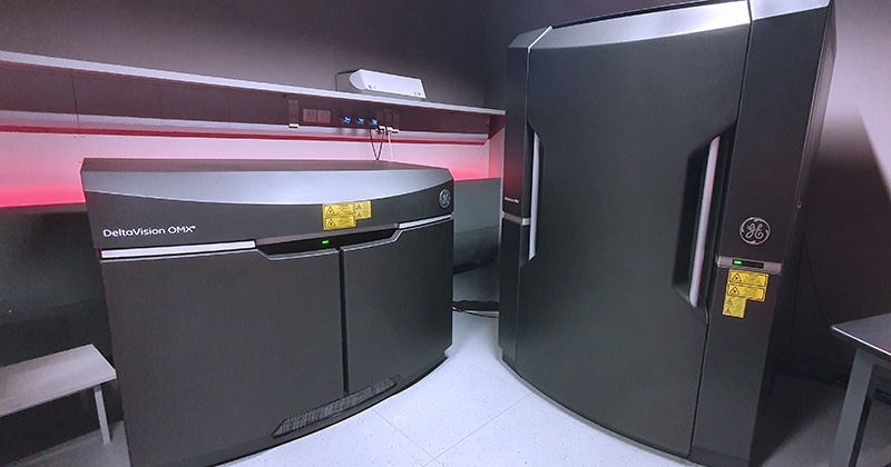
The DeltaVision OMX imaging platform is an advanced multi-mode, super-resolution inverted microscope system which offers super-resolution imaging using 3D Structured Illumination (3D-SIM) and Dense Stochastic Sampling Imaging (DSSI) – Localization Microscopy. The Blaze SIM Module offers dynamic high speed 3D-SIM suitable for live cell super-resolution imaging. Blaze incorporates a proprietary, ultra-fast, structured illumination module, advanced optical platform design and the latest high-speed camera technologies. The system can be used also for super-fast widefield acquisition and photo-kinetic experiments (FRAP).
Methods
Illumination
lamp for transmitted light
InsightSSI (Lumencore) illumination module – for WF imaging; standard shutters, open/close time ~ 1.8 ms excitation lasers – for 3D SIM, TIRF and localization; high speed galvanometer controlled shutters, open/close time ~ 200 μs
3D-SIM resolution at different wavelengths
| Laser | Type | Power (mW) | Expected XY resolution | Expected Z resolution |
| 405 nm | diode | 100 ± 10 | 110 ± 5 nm | 340 ± 10 nm |
| 445 nm | diode | 100 ± 5 | 115 ± 5 nm | 340 ± 10 nm |
| 488 nm | OPSL | 100 ± 4 | 120 ± 5 nm | 340 ± 10 nm |
| 514 nm | OPSL | 100 ± 6 | 120 ± 5 nm | 350 ± 10 nm |
| 568 nm | OPSL | 100 ± 4 | 135 ± 5 nm | 350 ± 10 nm |
| 642 nm | diode | 110 ± 10 | 160 ± 5 nm | 380 ± 20 nm |
Objectives
PLAN APO N 60x/1.42 OIL; FWD 0.15; CG 0.17 | BF, POL, DIC (optimized)
U APO N 60x/1.49 CORR OIL TIRF; FWD 0.1; CG 0.13-0.19 & 23/37°C | BF, POL, DIC
U APO N 100x/1.49 CORR OIL TIRF; FWD 0.1; CG 0.13-0.19 & 23/37°C | BF, POL, DIC
U PLAN S APO 60x/1.3 CORR SILICONE; FWD 0.3; CG 0.15-0.19 & 23/37°C | BF, POL, DIC
InsightSSI excitation filters (for widefield applications only)
DAPI 395.5/29
FITC 477/32
mCherry/Alexa Fluor 568 571/19
Cy5 645.5/15
CFP 438/24
YFP 512.5/15
Emission filters
DAPI 435.5/31
FITC 528/48
mCherry/Alexa Fluor 568 609/37
Cy5 683/40
CFP 477.5/35
YFP 541/22
Cameras
4x pco.edge 5.5 sCMOS, Readout speeds 95 MHz, 286 Mhz, 15 bit, pixel size: 6.5 μm camera, 80 nm 60x, 48 nm 100x
Stage
XYZ nanomover and fast Z-piezo
Additional equipment
antivibration table
sample holders:
Software
OMX Acquisition control software running on OMX Master computer under Windows 7
SoftWoRx – image reconstruction, deconvolution and analysis running on the SI workstation running under the CentOS v6 (Linux)
Dense Stochastic Sampling Imaging (DSSI) algorithm for localization microscopy images
Location: room no. 0.174 (Red)
Phone: ext. 2433
Booking: Calpendo („OMX“)
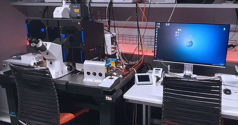
The Elyra 7 is a motorized inverted microscope equipped with a super-resolution system that uses a specialized lattice illumination pattern for 3D structured illumination (SIM) images and single-molecule localization methods (SMLM) like STORM and PALM. The lattice illumination allows for faster, gentler, and deeper imaging of live samples, and can reach up to 255 FPS during time-lapse acquisition. The unit has an incubation chamber for live imaging with controlled CO2 and temperature. The signal detection is achieved through two sCMOS cameras which enable simultaneous detection of two different fluorophores.
| Methods | Lattice SIM, SIM2 Single Molecule Localization Microscopy (3D PALM / dSTORM) TIRF Apotome (optical sectioning) mode Transmitted light imaging Brightfield and Nomarski contrast (DIC) (automatic) Time-lapse experiments (live-cell imaging) Multi-positions experiments |
| Illumination | Halogen 12 V/100W lamp – for transmitted light Metal halide HXP 120 V lamp – for fluorescence Excitation lasers (for TIRF, SIM, SMLM): – 405 nm diode, 50 mW – 488 nm OPSL, 500 mW – 561 nm OPSL, 500 mW – 642 nm OPSL, 500 mW |
| Objectives | EC Plan-Neofluar 10x/0.3 M27 DRY | BF, POL C-Apochromat 40x/1.2 Water Corr M27 | BF C-Apochromat 63x/1.2 Water Corr M27 | BF, DIC α Plan-Apochromat 63x/1.46 Oil Corr M27 | BF, TIRF α Plan-Apochromat 100x/1,46 Oil DIC M27 | BF, DIC, TIRF |
| Eyepiece filtercubes | Laser Blocking Filter for 405/488/561/642 named: – DAPI – FITC – mCherry – Cy5 Simultaneous two-channel viewing: – GFP + mCherry – GFP + Cy5 Sequential four-channel viewing: – DAPI and mCherry + GFP and Cy5 – DAPI and GFP + mCherry and Cy5 |
| Cameras | DuoLink for a parallel imaging on two cameras: – 2x pco.edge sCMOS (version 4.2 CL HS) – thermal stabilization at 0°C / +5 °C / +7 °C – 16-bit depth – high resolution: 2048 x 2048 px – pixel size 6.5 x 6.5 µm – quantum efficiency up to 82% – exposure times from 100 µs to 20 s – maximum frame rate 100 fps |
| Superresolution modules | Lattice SIM, SIM2 Single Molecule Localization Microscopy (3D PALM / dSTORM) |
| Stage | XY Scanning Stage Piezo 130 x 100 (D): – Movement range 130 mm x 100 mm (adjustable) – Max speed 100 mm/s – 0.2 μm resolution – Repeatability +/- 1 μm – Absolute accuracy +/- 5 μm Z-piezo insert: – Movement range at least 100 μm – < 5 nm resolution – Suitable for: Standard microscopic slip: 76 x 26 mm, Petri dish 30 – 35 mm in diameter, LabTek II chamber, 55 x 24 mm |
| Additional equipment | Antivibration table 120 x 90 cm Definite Focus – holding focus to compensate Z drift Dark incubation unit (CO2, temperature < 40°C, humidity) – Imaging chamber for Z-piezo inserts PC for storage and data analysis |
| Software | ZEN 3.0 (black edition) 64-bit Modules: – Lattice SIM2/SIM2 Apotome – SMLM (PALM/dSTORM) module – 3D-PALM – Measurement – Multi Channel – Panorama – Manual Extended Focus – Image Analysis – Time Lapse – Tiles & Positions – Z-stack – Extended Focus – Autofocus – Colocalization – Connect Entry (ZEN blue edition) |
| Location | room no. 0.171 (Orange) |
| Phone | ext. 3171 |
| Booking | Calpendo („Elyra 7“) |
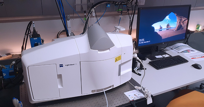
Z.1 light sheet fluorescence microscope allows fast, gentle multi-view imaging of large specimens. The microscope enables to record the development of living specimens and to deliver exceptionally high information content which includes the inner structures. To image deep into fixed non-transparent biological samples, it is necessary to apply a tissue clearing protocol (e.g. Scale, FocusClear, CUBIC).
| Methods | Fluorescence imaging with lightsheet optical sectioning Dual-site & multi-field scanning, processing at ZEN Black software Time-lapse experiments Multi-positions experiment |
| Illumination | 405nm Solid-state laser, 50mW 445nm Solid-state laser, 25mW 488nm Solid-state laser, 30mW 561nm Solid-state laser, 20mW 638nm Solid-state laser, 75mW Infrared LED light source for trasmitted light |
| Ilumination objectives | Objective Lightsheet Z.1 5x/0.1 Objective Lightsheet Z.1 10x/0.2 |
| Detection objectives | Dry objective Lightsheet Z.1 5x/0.16 Immersion objective Lightsheet Z.1 10x/0.5 Immersion objective Lightsheet Z.1 20x/1.0 Immersion objective Lightsheet Z.1 40x/1.0 Objective Clr Plan-Apochromat 20x/1.0 Corr nd=1,38 (suited for clearing media with RI 1.38, e.g. Scale) Objective Clr Plan-Neofluar 20x/1.0 Corr nd=1.45 (suited for clearing media with RI 1.45, e.g. FocusClear) |
| Laser blocking filters | 405/488/561 tripple excitation dichroic 405/488/561/640 quad excitation dichroic 405/488/640 tripple excitation dichroic 445/561 dual excitation dichroic |
| Emission filter sets | DAPI-GFP (SBS LP 490; Em: BP 420-470 + BP 505-545) DAPI-Cy3 (SBS LP 510; Em: BP 420-470 + BP 575-615) GFP-Cy3 (SBS LP 560; Em: BP 505-545 + BP 575-615) GFP-mCherry (SBS LP 560; Em: BP 505-545 + LP 585) GFP-DRAQ5 (SBS LP 560; Em: BP 505-545 + LP 660) GFP narrow-mCherry (SBS LP 560; Em: BP 505-530 + LP 585) |
| Cameras | Camera PCO.Edge 5.5 (sCMOS) – 2 pieces; 6,5 µm pixel |
| Additional equipment | Incubation unit (CO2, temperature, humidity) Antivibration table for SPIM microscopy PC for storage and data analysis Stereomicroscope for sample preparation and system maintenance (Stemi 305 EDU Microscope Set) |
| Software | ZEN Black system 2014 for Lightsheet Z.1 |
| Location | room no. 0.173 (Yellow) |
| Phone | ext. 2471 |
| Booking | Calpendo („Z1“) |

ScanR high content screening system is built on the inverted widefield microscope Olympus IX83. The system is designed for fully automated image acquisition and data analysis of biological samples. ScanR can handle many different specimen formats, including multi-well plates, slides and custom-built arrays. The hardware autofocus maintains focus stability within the entire specimen. The acquisition is followed by complex image analysis and advanced data evaluation including utilization of pre-trained AI models. Highly sensitive camera enables low fluorescence intensity applications.
| Methods | High-content imaging (widefield) |
| Illumination | Lumencore SPECTRA X IR LED light source for fluorescence (peaks: 377, 438, 475, 510, 555, 575, 635 and 730 nm) Lamp for transmitted light |
| Objectives | UPLSAPO 10x/0.4 DRY; FWD 3.1; CG 0.17 UPLXAPO 20x/0.8 DRY CORR; FWD 0.6; CG 0 – 2 UPLXAPO 40x/0.95 DRY CORR; FWD 0.18; CG 0.11 – 0.23 UPLXAPO 60x/1.42 OIL; FWD 0.15; 0.15 |
| Excitation LED source | UV – DAPI – 377/54 nm (700 mW) Blue – CFP – 438/29 nm (700 mW) Cyan – GFP (FITC) – 475/28 nm (300 mW) Green – TRITC (Cy3) – 555/28 nm (600 mW) Red – Cy5 – 635/22 nm (200 mW) NIR – Cy7 – 730/40 nm (300 mW) |
| Emission filtercubes | PENTA – 5-band dichroic: DAPI/FITC/Cy3/Cy5/Cy7 (409/493/573/652/759 nm) DIC |
| Emission filters | DAPI (432/36) FITC (GFP) (515/30) Cy3 (TRITC) (595/31) Cy5 (685/40) Cy7 (809/81) |
| Cameras | sCMOS camera Hamamatsu ORCA-Fusion BT, 6.5 µm pixel, QE >95%, cooled to -8°C, resolution 2304 x 2304 pixels |
| Stage | Motorized microscope stage xyz |
| Aditional equipment | DFC hardware autofocus |
| Software | ScanR acquisition ScanR analysis |
| Location | room no. 0.175 (Blue) |
| Phone | ext. 2750 |
| Booking | Calpendo („ScanR-1“) |

ScanR high content screening system is built on the inverted widefield microscope Olympus IX83. The system is designed for fully automated image acquisition and data analysis of biological samples. ScanR can handle many different specimen formats, including multi-well plates, slides and custom-built arrays. The hardware autofocus maintains in-focus stability within the entire specimen. The acquisition is followed by complex image analysis and advanced data evaluation including utilization of pre-trained AI models. Highly sensitive camera enables low fluorescence intensity applications.
| Methods | High-content imaging (widefield) |
| Illumination | Lumencore Spectra X LED light source for fluorescence (peaks: 395, 440, 470, 510, 550, 575 a 640 nm) Lamp for transmitted light |
| Objectives | UPLSAPO 10x/0.4 DRY; FWD 3.1; CG 0.17 UPLXAPO 20x/0.8 DRY CORR; FWD 0.6; CG 0 – 2 UPLXAPO 40x/0.95 DRY CORR; FWD 0.18; CG 0.11 – 0.2 UPLXAPO 60x/1.42 OIL; FWD 0.15; 0.15 |
| Excitation LED source | UV – DAPI 395/25 nm (295mW) Blue – CFP 440/20 nm (256mW) Cyan – FITC 470/24 nm (196mW) Teal – YFP 510/25 nm (52mW) Green – Cy3 550/15 nm (260mW) Red – Cy5 640/30 nm (231mW) |
| Emission filtercubes | D/F/Cy3/Cy5: 4-band dichroic: DAPI/FITC/Cy3/Cy5 (409/493/573/652 nm) CFP/YFP/mCherry: 3-band dichroic: CFP/YFP/mCherry (459/526/596 nm) BF |
| Emission filters | DAPI (433/24) CFP (482/25) FITC (GFP) (520/35) YFP (544/24) Cy3 (TRITC) (600/37) mCherry (641/75) Cy5 (680/42) |
| Cameras | sCMOS camera Hamamatsu ORCA-Fusion BT, 6.5 µm pixel, QE >95%, cooled to -8°C, resolution 2304 x 2304 pixels |
| Stage | Motorized microscope stage xyz |
| Aditional equipment | Hardware autofocus |
| Software | ScanR acquisition ScanR analysis |
| Location | room no. 0.175 (Blue) |
| Phone | ext. 2750 |
| Booking | Calpendo („ScanR-2“) |
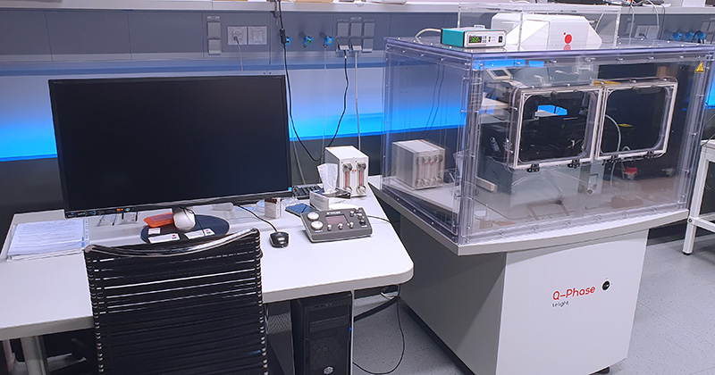
Q-PHASE is a unique instrument for quantitative phase imaging (QPI). It is based on a robust and fully motorized inverted transmission microscope platform. Multiple imaging modes are integrated such as fluorescence, simulated DIC, high-pass filter and brightfield. It is a perfect tool for long-term label-free imaging of living cells. Sophisticated user-friendly software is available for microscope control, image acquisition and subsequent cell segmentation and highly advanced data mining and analysis.
| Methods | Quantitative phase imaging in TL mode Additional fluorescence imaging Time-lapse experiments Multi-position experiments QPI image advanced analysis |
| Illumination | Lumencore Spectra X LED light source for fluorescence Transmitted light source halogen lamp EKE 21V |
| Objectives | Nikon PLAN ACHROMAT 4x/0.1 DRY; FWD 30 | BF, QPhase, Hologram, POL, DIC, High Pass Filter (PH) Nikon PLAN FLUOR 10x/0.3 DRY; FWD 16 | BF, QPhase, Hologram, POL, DIC, High Pass Filter (PH) Nikon PLAN FLUOR 20x/0.5 DRY; FWD 2.1 | BF, QPhase, Hologram, POL, DIC, High Pass Filter (PH) Nikon PLAN APO 40x/0.95 DRY, FWD 0.17-0.25 | BF, QPhase, Hologram, POL, DIC, High Pass Filter (PH) Nikon PLAN APO 60x/1.4 OIL, FWD 0.13| BF, QPhase, Hologram, POL, DIC, High Pass Filter (PH) |
| Filter set | DAPI FITC/Texas Red |
| Spectra X excitation filters | DAPI 390/22 CFP 434/21 GFP 470/24 FITC 494/20 YFP 511/16 DsRED 555/28 Texas Red 575/25 |
| Dichronic mirrors | QPl-Fluorescence Beam Combiner SP 635 (limits detection of emission wavelengths below 635 nm) 436/514/604 nm tripple band dichroic DAPI/FITC/Texas Red (transmission bands Tavg > 90%: 446-468, 520-540, 614-642) 440/520 nm dual band dichroic CFP/YFP (transmission bands Tavg > 90%: 449-483, 530-569) 493/574 nm dual band dichroic GFP/DsRed (transmission bands Tavg > 90%: 500-529, 584-679) |
| Emission filter wheel | DAPI 447/60 CFP 472/30 GFP 514/30 FITC 531/22 YFP 542/27 DsRed 617/73 Texas Red 624/40 |
| Cameras | XIMEA MR4021MC – monochromatic CCD camera for TL; 7,4 µm pixel Andor Zyla 5.5 – monochromatic sCMOS camera for fluorescence; 6,5 µm pixel |
| Aditional equipment | incubation unit (CO2, temperature, humidity) |
| Software | SophiQ software |
| Location | room no. 0.175 (Blue) |
| Phone | ext. 2750 |
| Booking | Calpendo („QPI“) |
| Software | FiJi ImageJ Horizon client provides access to Virtual Desktops equipped with various proprietary and free software packages for image processing and analysis. |
| Hardware | CPU 16 core E5-2640, 2.6 GHz (PassMark – 21311) RAM 128 GB GPU GeForce GTX 1080 8 GB LAN 10 Gb/s |
| Location | room no. 0.158 (Analysis) |
| Phone | ext. 2258 |
| Booking | Calpendo („Analysis 1“) |

| Software | FiJi ImageJ Horizon client provides access to Virtual Desktops equipped with various proprietary and free software packages for image processing and analysis. |
| Hardware | CPU 8 core i7-7820X, 3.6 GHz (PassMark – 18920) RAM 128 GB GPU GeForce GTX 1080 Ti 11 GB LAN 10 Gb/s |
| Location | room no. 0.158 (Analysis) |
| Phone | ext. 2258 |
| Booking | Calpendo („Analysis 2“) |

| Software | FiJi ImageJ Horizon client provides access to Virtual Desktops equipped with various proprietary and free software packages for image processing and analysis. |
| Hardware | CPU 4 core E3-1270, 3.8 GHz (PassMark – 11548) RAM 32 GB GPU GeForce GTX 1080 Ti 11 GB LAN 1 Gb/s |
| Location | room no. 0.158 (Analysis) |
| Phone | ext. 2258 |
| Booking | Calpendo („Analysis 3“) |

| Software | FiJi ImageJ Horizon client provides access to Virtual Desktops equipped with various proprietary and free software packages for image processing and analysis. |
| Hardware | CPU 24 core e5-2620, 2.4 GHz (PassMark – 16139) RAM 128 GB GPU GeForce GTX 1080 8 GB LAN 10 Gb/s |
| Location | room no. 0.158 (Analysis) |
| Phone | ext. 2258 |
| Booking | Calpendo („Analysis 4“) |
A high-performance workstation designed primarily for applications with demanding requirements for computing resources and RAM, such as image stitching, processing large datasets from light-sheet or spinning disk microscopes, and deep learning applications.
| Software | Imaris – Core and Measurement Pro + floating licenses: Vantage, Coloc, XT (4 of each) Track, Lineage, Cells Viewer, Filament, Batch (2 of each) Imaris Stitcher NIS-Elements with Machine Learning Module Anaconda (Python) FiJi ImageJ |
| Hardware | CPU 32-core RAM 1024 GB GPU Nvidia A40 24GB |
| Booking | Calpendo („01 – Imaris, Stitcher, NIS-E (Gold)“) |
| Access | Virtual Desktop Infrastructure („01 – Imaris, Stitcher, NIS-E (Gold)“) |
A high-performance workstation designed primarily for applications requiring significant computing resources and RAM, such as image stitching, processing large datasets from light-sheet or spinning disk microscopes, and deep learning. The Huygens deconvolution software on this virtual machine leverages a graphics processor (NVIDIA A40, 44GB) for computations and includes an option for light-sheet data deconvolution.
| Software | Huygens Professional – Widefield & Brightfield, Confocal, STED 3D, Light Sheet, Bleaching Corrector, Chromatic Aberration Corrector, Crosstalk corrector, Tile Stitching, Light Sheet Fuser, Object Stabilizer, PSF Distiler, Workflow Processor, Batch Feeder NIS-Elements 7.0 beta with Machine Learning Module Anaconda (Python) FiJi ImageJ |
| Hardware | CPU 32-core RAM 1024 GB GPU Nvidia A40 44GB |
| Booking | Calpendo („02 – Huygens-GPU, NIS-E (Gold)“) |
| Access | Virtual Desktop Infrastructure („02 – Huygens-GPU, NIS-E (Gold)“) |
A powerful workstation specifically designed for applications utilizing software packages on Linux. This machine is ideal for tackling complex tasks such as the analysis and registration of volumetric data, cell tracking in entire developing embryos, and training or inferencing deep learning networks for image or volume segmentation and classification tasks. Its capabilities are especially enhanced by the power of GPU computation. Specialized training is required to operate this virtual machine.
| Software | Anaconda (Python) Other software packages can be installed on demand |
| Hardware | CPU 8-core RAM 256 GB GPU Nvidia A40 16GB |
| Booking | Calpendo („03 – Linux (Silver)“) |
| Access | Virtual Desktop Infrastructure („03 – Linux (Silver)“) |
Powerful workstation for image analysis. In addition to the standard ImageJ and Python packages, it also offers Imaris software and NIS-Elements software.
| Software | Imaris – Core and Measurement Pro + floating licenses: – Vantage, Coloc, XT (4 of each) – Track, Lineage, Cells Viewer, Filament, Batch (2 of each) NIS-Elements with Machine Learning Module Anaconda (Python) FiJi ImageJ |
| Hardware | CPU 32-core RAM 256 GB GPU Nvidia A40 24GB |
| Booking | Calpendo („05 – Imaris, NIS-E (Silver)“) |
| Access | Virtual Desktop Infrastructure („05 – Imaris, NIS-E (Silver)“) |
Standard workstation for image analysis. In addition to the standard ImageJ and Python packages, it also offers Imaris software, NIS-Elements software. ZEN Black 3.0 software enables processing of SIM and SMLM super-resolution data from Elyra 7 microscope.
| Software | Imaris – Core and Measurement Pro + floating licenses: – Vantage, Coloc, XT (4 of each) – Track, Lineage, Cells Viewer, Filament, Batch (2 of each) ZEN Black 3.0 – Deconvolution, Lattice SIM & SIM2 Processing, Localization microscopy processing NIS-Elements with Machine Learning Module Anaconda (Python) FiJi ImageJ |
| Hardware | CPU 12-core RAM 128 GB GPU Nvidia A40 16GB |
| Booking | Calpendo („06 – Imaris, ZEN-SR, NIS-E“) |
| Access | Virtual Desktop Infrastructure („06 – Imaris, ZEN-SR, NIS-E“) |
Standard workstation for image analysis. In addition to the standard ImageJ and Python packages, it also offers Imaris software and NIS-Elements software. Matlab programming and numeric computing platform provides another tool for data analysis, signal and image processing. Imaris software can be bridged with Matlab via Imaris XT module.
| Software | Imaris – Core and Measurement Pro + floating licenses: – Vantage, Coloc, XT (4 of each) – Track, Lineage, Cells Viewer, Filament, Batch (2 of each) Matlab – Image processing toolbox, Signal processing toolbox, Statistical toolbox, Optimization toolbox, Curve fitting toolbox NIS-Elements with Machine Learning Module Anaconda (Python) FiJi ImageJ |
| Hardware | CPU 12-core RAM 128 GB GPU Nvidia A40 16GB |
| Booking | Calpendo („07 – Imaris, Matlab, NIS-E“) |
| Access | Virtual Desktop Infrastructure („07 – Imaris, Matlab, NIS-E“) |
Standard workstation for image analysis. In addition to the standard ImageJ and Python packages, it also offers Imaris software and NIS-Elements software. Matlab programming and numeric computing platform provides another tool for data analysis and image processing. Imaris software can be bridged with Matlab via Imaris XT module.
| Software | Imaris – Core and Measurement Pro + floating licenses: – Vantage, Coloc, XT (4 of each) – Track, Lineage, Cells Viewer, Filament, Batch (2 of each) Matlab – Image processing toolbox, Statistical toolbox NIS-Elements with Machine Learning Module Anaconda (Python) FiJi ImageJ |
| Hardware | CPU 12-core RAM 128 GB GPU Nvidia A40 16GB |
| Booking | Calpendo („08 – Imaris, Matlab, NIS-E“) |
| Access | Virtual Desktop Infrastructure („08 – Imaris, Matlab, NIS-E“) |
Standard workstation for image analysis. In addition to the standard ImageJ and Python packages, it also offers Imaris software and Arivis software.
| Software | Imaris – Core and Measurement Pro + floating licenses: – Vantage, Coloc, XT (4 of each) – Track, Lineage, Cells Viewer, Filament, Batch (2 of each) Arivis 3.1.3 Anaconda (Python) FiJi ImageJ |
| Hardware | CPU 12-core RAM 128 GB GPU Nvidia A40 16GB |
| Booking | Calpendo („09 – Imaris, Arivis“) |
| Access | Virtual Desktop Infrastructure („09 – Imaris, Arivis“) |
A high-performance workstation designed primarily for applications with demanding requirements for computing resources and RAM, such as image stitching, processing large datasets from light-sheet or spinning disk microscopes, and deep learning applications. The Huygens deconvolution software on this virtual machine utilizes a CPU (AMD EPYC 7713, 64-core, 2.0GHz) for computations and includes an option for spinning disk data deconvolution.
| Software | Huygens Professional – Widefield & Brightfiled , Confocal, Spinning Disk & SoRa, STED 3D, Bleaching Corrector, Chromatic Aberration Corrector, Object Stabilizer, PSF Distiler, Movie Maker, Object Analyzer, Object Tracker, Colocalization Analyzer with RBNCC option, Workflow Processor, Batch Feeder ZEN Blue 3.4 – Deconvolution, Light sheet Processing, MultiView & Dual Side Fusion NIS-Elements with Machine Learning Module Anaconda (Python) FiJi ImageJ |
| Hardware | CPU 64-core RAM 512 GB GPU Nvidia A40 24GB |
| Booking | Calpendo („10 – Huygens-CPU, ZEN-LS, NIS-E (Gold)“) |
| Access | Virtual Desktop Infrastructure („10 – Huygens-CPU, ZEN-LS, NIS-E (Gold)“) |
Standard workstation for image analysis. CellSense Dimensions software and ScanR Analysis software from Evident (Olympus) are available. Workstation also offers AI deep learning module that works together with both software packages and enables deep learning of new AI models for segmentation.
| Software | ScanR Analysis software with AI Deep Learning Module cellSense Dimension – Deconvolution, AI Deep Learning Module Anaconda (Python) FiJi ImageJ |
| Hardware | CPU 12-core RAM 128 GB GPU Nvidia A40 16GB |
| Booking | Calpendo („11 – ScanR, cellSense“) |
| Access | Virtual Desktop Infrastructure („11 – ScanR, cellSense“) |
Standard workstation for image analysis. It offers ZEN software for working with Zeiss specific file formats. In collaboration with the Flow Cytometry Core Facility, it also enables analysis of image cytometry data from the Image Stream flow cytometer using IDEAS software with a machine learning module.
| Software | IDEAS software with Machine Learning Module ZEN – Time Series, Multi-Channel, Measurement Anaconda (Python) FiJI ImageJ |
| Hardware | CPU 12-core RAM 64 GB GPU Nvidia A40 8GB |
| Booking | Calpendo („12 – Ideas ML, ZEN“) |
| Access | Virtual Desktop Infrastructure („12 – Ideas ML, ZEN“) |
Standard workstation for image analysis. It offers the analysis of FLIM and FCS data from the Stellaris 8 microscope. In collaboration with the Flow Cytometry Core Facility, it also enables the analysis of image cytometry data from the Image Stream flow cytometer using the IDEAS and Amnis AI software packages.
| Software | IDEAS software Amins AI Software Leica LAS X – FLIM, Phasor FLIM, FCS Anaconda (Python) FiJI ImageJ |
| Hardware | CPU 12-core RAM 64 GB GPU Nvidia A40 8GB |
| Booking | Calpendo („13 – Ideas AI, LAS X“) |
| Access | Virtual Desktop Infrastructure („13 – Ideas AI, LAS X“) |
Workstation operated in collaboration with the Flow Cytometry Core Facility, that provides access to FlowJo software for flow cytometry data analysis. It can be used for more sophisticated analysis of FCS files exported from ScanR Analysis software. In addition, R programming language is also available.
| Software | FlowJo software R software |
| Hardware | CPU 8-core RAM 32 GB GPU not dedicated |
| Booking | Calpendo („14 – FlowJo“) |
| Access | Virtual Desktop Infrastructure („14 – FlowJo“) |
Huygens Core compute engine is available on the powerful server with two GPUs, that allows simultaneous batch deconvolution of files up to 100 GB. The Huygens Remote Manager (HRM) interface allows user to configure and run deconvolution batch jobs.
| Software | Huygens Core – Widefield & Brightfiled, Confocal, Spinning Disk & SoRa, Multi Photon, STED 3D, Light Sheet, Object Stabilizer, Chromatic Aberration Corrector |
| Hardware | CPU 24-core RAM 1760 GB GPU 2 x Nvidia RTX A5000 24GB |
| Booking | Not needed |
| Access | Huygens Remote Manager |