Institute of Molecular Genetics of the Czech Academy of Sciences

Inverted microscope DMi8 with confocal head Leica TCS SP8 is a confocal laser scanning microscope with full transmitted light equipment including transmitted light PMT and Nomarski contrast (DIC) available for all objectives. Leica TCS SP8 it is a dichroic mirror-based system, using interference filters for light filtering and beam-splitting. The confocal head is equipped with two fluorescence PMT detectors and three highly sensitive HyD detectors. The laser power is modulated by acousto-optical tunable filter (AOTF).
| Methods | Confocal scanning fluorescence imaging Transmitted light imaging Brightfield and Nomarski contrast (DIC) (manual) Tile-scans & merge in LAS X software Time-lapse experiments Multi-positions experiment Photo-kinetic experiment FRET (SE or Acceptor photobleaching) |
| Illumination | 405 nm diode laser, 50mW 488 nm solid-state laser, 20 mW 552 nm solid-state laser, 20 mW 638 nm solid-state laser, 30 mW HLX 100 W halogen lamp for transmitted light HXP 120W/45C VIS Hg lamp – Leica EL6000 for fluorescence |
| Objectives | HC PL FLUOTAR 10x/0.30 DRY; FWD 11.0 | BF, POL, DIC HC PL FLUOTAR 10x/0.30 DRY; FWD 11.0 | BF, POL, DIC HC PL APO 20x/0.75 IMM CORR CS2; FWD 0.66 | BF, POL HC PL APO 40x/1.30 OIL CS2; FWD 0.24; CG 0.17 | BF, POL, DIC HCX PL APO 63x/1.3 GLYC CORR 37°C; FWD 0.28; CG 0.14-0.18 | BF, POL HC PL APO 63x/1.40 OIL CS2; FWD 0.14; CG 0.17 | BF, POL, DIC |
| Eyepiece filtercubes | DAPI (A) (Ex: 360/40, Em: LP 425) FITC (I3) (Ex: 470/40; DM 510; Em: LP 515) RHOD (N2.1) (Ex: 537/45; DM 580; Em: LP 590) Cy5 (Y5) (Ex: 620/60; DM 660; Em: 700/75) CFP (Ex: 436/20; DM 455; Em: 480/40) |
| Detectors | 2x photomultiplier tube (PMT) 3x supersensitive hybrid detectors (HyD) 1x transmitted light detector |
| Confocal head | Acousto-optical tunable filter (AOTF) Low incident angle dichroic beam splitters Standard scanner (1-1800 Hz line frequency) Maximum scanner resolution 8192×8192 pixels Hardware zoom 0.75x-48x |
| Dichroic mirrors | 488/552/638 nm triple excitation dichroic 488/552 nm dual excitation dichroic Substrate RT 15/85 |
| Stage | Motorized microscope stage with Super Z-galvo scanning insert for fast and precise Z movement |
| Aditional equipment | Antivibration table |
| Software | LAS X |
| Location | room no. 0.172 (Green) |
| Phone | ext. 3172 |
| Booking | Calpendo (“SP8” ) |

The inverted microscope DMi8 with laser scanning confocal head Leica TCS SP8 is a confocal laser scanning microscope with full transmitted light equipment including transmitted light PMT and Nomarski contrast (DIC) available for all objectives. The confocal head is equipped with two fluorescence PMT detectors and three highly sensitive HyD detectors with time resolved gating function useful e.g. for removal of auto-fluorescence. The system has a wide-range white light laser with pulse picker (WLL PP) which allows for very versatile illumination tailored for any fluorophore. The combination of white light laser, AOBS and spectral sliders on detector cascade allows the fine tuning of excitation laser line selection and precise definition of emission spectra. Confocal head light path is equipped with optical image rotation. Stability for long-term live cell imaging experiments is ensured by a hardware autofocus system (AFC) and optional OKOLAB stage-top incubation chamber, which provides temperature, CO2, and humidity control.
| Methods | Confocal scanning fluorescence imaging Brightfield and Nomarski contrast (DIC) (automated) Tile-scans & merge in LAS X software Time-lapse experiments Multi-positions experiment |
| Illumination | Lamp for transmitted light: Leica EL6000 with HXP 120W/45C VIS Hg lamp – for fluorescence Excitation lasers: Pulsed white light laser (WLL2) – 470-640 nm with 1nm step, 8 parallel laser lines, 1.5 mW per each Continuous 405 nm DMOD Flexible laser – 50 mW |
| Objectives | HC PL FLUOTAR 10x/0.30 DRY; FWD 11.0 | BF, POL, DIC HC PL APO 20x/0.75 IMM CORR CS2; FWD 0.66 | BF, POL HC PL APO 40x/1.30 OIL CS2; FWD 0.24; CG 0.17 | BF, POL, DIC HC PL APO 63x/1.40 OIL CS2; FWD 0.14; CG 0.17 | BF, POL, DIC HC PL APO 100x/1.4 OIL STED WHITE CS2; FWD 0.13; CG 0.17 | BF, POL, DIC (OPTIONAL – UPON REQUEST) HC PL APO 86x/1.20 W CS2; FWD 0.30 mm| BF, POL, DIC |
| Eyepiece filtercubes | DAPI (A) (Ex: 360/40; DM 400; Em: LP 425) FITC (I3) (Ex: 470/40; DM 510; Em: LP 515) RHOD (N2.1) (Ex: 537/45; DM 580; Em: LP 590) Cy5 (Y5) (Ex: 620/60; DM 660; Em: 700/75) |
| Detectors | 2x photomultiplier tube (PMT) 400-800 nm 3x supersensitive hybrid detectors (HyD) 400-720 nm 1x transmitted light detector |
| Confocal head | Acousto-optical tunable filter (AOTF) Acousto-optical beam splitter (AOBS) Optical rotation Standard scanner (1-1800 Hz line frequency) Maximum scanner resolution 8192×8192 pixels Spectrally tunable detection Hardware zoom 0.75x-48x |
| Stage | Motorized microscope stage with Super Z-galvo scanning insert for fast and precise Z movement HW autofocus control |
| Aditional equipment | Antivibration table Incubation chamber Okolab (CO2, temperature, humidity) |
| Software | LAS X 64bit |
| Location | room no. 0.172 (Green) |
| Phone | ext. 3172 |
| Booking | Calpendo (“SP8 WLL” ) |
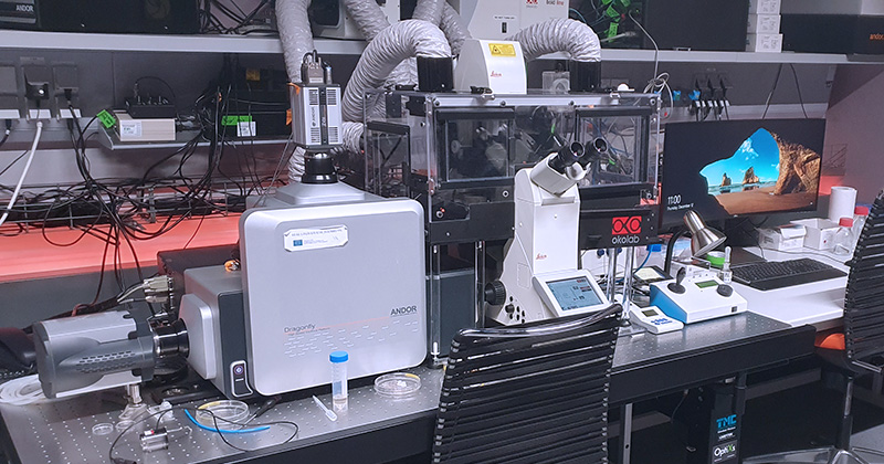
Dragonfly is a fully motorized multi-modal imaging platform with confocal and widefield modes. The core of the microscope is the confocal spinning disk, which combined with high-speed sCMOS and high-sensitive EMCCD cameras provides the system with exceptional speed and sensitivity. The low phototoxicity and photobleaching is ideal for live or delicate specimens. Dragonfly is 10-20 times faster than a traditional laser scanning confocal, which makes it optimal for imaging of fast dynamic events in live cell or for high throughput experiments. Photomanipulation experiments can be performed with Mosaic, a tool that allows for simultaneous illumination of multiple regions of interest of flexible shape and size. The semi-automated microinjector suited for adherent cells microinjection is also available. System is equipped with Super Resolution Radial Fluctuations super-resolution method.
| Methods | Confocal spinning disk fluorescence imaging Transmitted light imaging Brightfield and Nomarski contrast (DIC) Large tile-scans & merge in Fusion software Time-lapse experiments with HW autofocus Multi-positions experiment Photo-kinetic and opto-genetic experiment (limited software solution) Super Resolution Radial Fluctuations (SRRF) |
| Illumination | 405 nm solid-state laser, 200 mW 445 nm solid-state laser, 75 mW 488 nm solid-state laser, 150 mW 514 nm solid-state laser, 40 mW 561 nm solid-state laser, 100 mW 637 nm solid-state laser, 140 mW White LED source Leica DM for transmitted light CoolLED pE-300; 365 UV – white LED source for fluorescence 445 nm solid-state photo-manipulation laser for Mosaic, 1.3 W White LED source for Mosaic photo-manipulation (CoolLED pE-300; 365 UV) |
| Objectives | (HC PL APO 10x/0.40 DRY CS2; FWD 2.2; CG 0.17 | BF, POL, DIC) (OPTIONAL, UPON REQUEST) HC PL APO 20x/0.75 IMM CORR CS2; FWD 0.66 | BF, POL, DIC HC PL APO 40x/1.10 W CORR CS2; FWD 0.65; CG 0.14-0.18 | BF, POL, DIC HC PL APO 63x/ 1.20 W CORR CS2; FWD 0.3; CG 0.14-0.18 | BF, POL, DIC (OPTIONAL, UPON REQUEST) HCX PL APO 40x/1.25-0.75 OIL λBL; FWD 0.13; CG 0.17 | BF, POL HCX PL APO 63x/1.40-0.6 OIL λB; FWD 0.12; CG 0.17 | BF, POL, DIC HCX PL APO 63x/1.3 GLYC CORR 37°C; FWD 0.28; CG 0.14-0.18 | BF, POL HCX PL APO 100x/1.4-0.7 Oil CS; FWD 0.13; CG 0.17 | BF, POL, DIC |
| Dichroic mirrors | 405/488/561/640 nm Quad excitation dichroic 399-452/514/640 nm Triple excitation dichroic 405/488/561/685 nm Quad excitation dichroic |
| Eyepiece filtercubes | DAPI (Ex: 350/50; DC: 400; Em: 460/50) CFP/YFP (Ex: 435/25, 500/20; DC: 450; Em: 470/25, 535/20) FITC (Ex: 480/40; DC: 505; 527/30) RHOD (Ex: 546/10; DC: 560; 585/40) |
| Cameras | Zyla 4.2 PLUS sCMOS camera – 2048 x 2048 pix; 6,5 µm pixel iXon Ultra 888 EMCCD camera – 1024 x 1024 pix; 13 µm pixel |
| Dual camera switching mirror | LP 500 nm dichroic mirror (reflection to Zyla camera) LP 565 nm dichroic mirror (reflection to Zyla camera) 100% mirror (Zyla camera) 100% transmission dummy glass (iXon camera) |
| Zyla camera emission filter wheel | 450/50 nm bandpass filter (DAPI) 480/40 nm bandpass filter (CFP) 525/50 nm bandpass filter (GFP, A488, FITC) 540/30 nm bandpass filter (YFP) 600/50 nm bandpass filter (RFP, TRITC) 700/75 nm bandpass filter (Cy5) 405 to 445-515-561-730 quad emission filter (DAPI/CFP/YFP/mCherry/DyLight) Polarization filter |
| iXon camera emission filter wheel | 450/50 nm bandpass filter (DAPI) 480/40 nm bandpass filter (CFP) 525/50 nm bandpass filter (GFP, A488, FITC) 540/30 nm bandpass filter (YFP) 600/50 nm bandpass filter (RFP, TRITC) 620/60 nm bandpass filter (mCherry) 700/75 nm bandpass filter (Cy5) 405-488-561-640 quad emission filter (DAPI/GFP/RFP/Cy5) |
| Spare camera emission filter wheel | Polarization filter 725/40 nm bandpass filter 405-488-561-640 quad emission filter (DAPI/GFP/RFP/Cy5) 405 to 445-515-561-730 quad emission filter (DAPI/CFP/YFP/mCherry/DyLight) 445-515-561 tripple emission filter (CFP/YFP/RFP) 488-561 dual emission filter (GFP/RFP) 445-515 dual emission filter (CFP/YFP) |
| Microinjection | Microinjector FemtoJet joined with micromanipulator InjectMan NI 2 (Eppendorf) For volumes up to 100 pl Compressor to deliver the required pressure Integrated coarse and fine manipulator Work range: 20 mm per axis Automated and programmable axial injection movement |
| Aditional equipment | Okolab incubation unit including climate chamber (CO2, temperature, humidity) antivibration table photomanipulation module Mosaic (for FRAP, bleaching, uncaging, opto-genetics…) |
| Software | Fusion – acquisition software Imaris – visualization software Andor iQ3 – controlling Mosaic module |
| Location | room no. 0.171 (Orange) |
| Phone | ext. 3171 |
| Booking | Calpendo (“Dragonfly”) |

IXplore SpinSR SoRa is a fully motorized multimodal fluorescence microscope designed for gentle and rapid live-cell imaging. Built on the Evident IX83 platform, the system features a modular two-tier frame with an integrated focus adjustment for Köhler illumination and motorized objective turret that can accommodate up to six objectives. The system supports advanced imaging methods, including confocal spinning disk microscopy and super-resolution imaging based on optical reassignment technology. The system integrates cellSense software solution for multi-dimensional data acquisition, including z-stacks, multi-channel, tile-scans, time-lapse, and multi-position imaging. It also features scanR acquisition and analysis software for high-content screening. The microscope is equipped with drift focus correction (DFC) system to maintain z-axis stability during long term experiments.
| Methods | Confocal spinning disk with 50um pinholes (standard confocal) Confocal spinning disk with SoRa technology for enhanced resolution up to 120 nm Transmitted light imaging Large tile-scans & merge Time-lapse experiments with HW autofocus Multi-positions experiment DFC system based on IR laser for real-time imaging and precise Z-axis stabilization High-content screening with scanR software |
| Illumination | 405 nm solid-state laser, 50 mW 488 nm solid-state laser, 150 mW 561 nm solid-state laser, 100 mW 640 nm solid-state laser, 100 mW 785 nm solid-state laser, 100 mW Transmitted light: LED source IX3-LHLEDC CoolLED pE-300white for widefield fluorescence |
| Objectives | UPLXAPO 10X/0.40 DRY CS2; FWD 3.1 mm; CG 0.17 UPLXAPO 40X/0.95 IMM CORR CS2; FWD 0.18 mm; CG 0.11-0.23 UPLXAPO 60XO/1.42 OIL CS2; FWD 0.15 mm; CG 0.17 UPLXAPO 100XO/1.45 OIL CS2; FWD 0.13 mm; CG 0.17 |
| Dichroic mirrors | 405/488/561/640 nm Quad excitation dichroic 785 nm Single dichroic |
| Eyepiece filtercubes | DAPI (Ex: 350/50; DC: 400; Em: 460/50) FITC (Ex: 480/40; DC: 505; Em: 527/30) Cy3 (Ex: 546/10; DC: 560; Em: 585/40) |
| Cameras | ZHamamatsu ORCA-Fusion BT Type: sCMOS, global shutter Resolution: 2304 x 2304 pixels (5.2 MPix). Pixel Size: 6.5 x 6.5 µm Quantum Efficiency (QE): >95% at 550 nml |
| Dual camera switching mirror | LP 561 dichroic mirror: Directs emission to separate channels for simultaneous dual-camera imaging (e.g., GFP and RFP) Clear glass mirror: Allows full transmission to a single camera |
| Camera emission filter wheel | 2x 10-position wheels, each camera equipped with: DAPI (Ex: 405 nm) FITC (Ex: 488 nm) Cy3 (Ex: 561 nm) Cy5 (Ex: 640 nm) Cy7 (Ex: 785 nm) |
| Aditional equipment | Peacon incubation unit including climate chamber (CO2/O2, temperature, humidity) antivibration table |
| Software | CellSense acquisition software for confocal multimodal imaging ScanR Acquisition software for high-content screening ScanR Analysis software for high-content data analysis |
| Location | room no. 0.175 (Blue) |
| Phone | ext. 2750 |
| Booking | Calpendo (“SpinSR”) |